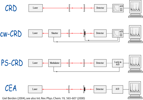
In 1988 O'Keefe and Deacon demonstrated a new direct absorption spectroscopic technique that can be performed with a pulsed light source and that has a significantly higher sensitivity than obtainable in `conventional' absorption spectroscopy. This so-called Cavity Ring Down (CRD) technique is based upon the measurement of the rate of absorption rather than the magnitude of absorption of a light pulse confined in a closed optical cavity with a high Q-factor. The advantage over normal absorption spectroscopy results from (i) the intrinsic insensitivity of the CRD technique to light source intensity fluctuations, and (ii) the extremely long effective path lengths (many kilometers) that can be realized in stable optical cavities. During the last decades research on various CRD detection schemes has exploded, as recently described in our review paper and book. A short overview can be found in our paper in American Laboratory.
In a typical CRD experiment a short light pulse is coupled into a stable optical cavity, formed by two highly reflecting plano-concave mirrors. The fraction of light entering the cavity on one side `rings' back and forth many times between the two mirrors. The time behavior of the light intensity inside the cavity I_{CRD}(t), can be monitored by the small fraction of light that is transmitted through the other mirror. If the only loss factor in the cavity is the reflectivity loss of the mirrors, one can show that the light intensity inside the cavity decays exponentially in time with a decay constant tau, the `ring down time', given by: tau = d/{c(|ln R|)}. Here d is the optical path length between the mirrors, c is the speed of light and R is the reflectivity of the mirrors. If R is close to unity, the approximation tau = d/c(1-R) can be made.
 If there is additional loss inside the cavity due to the presence of absorbing
and light scattering species, the light intensity inside the cavity will
still decay exponentially in time provided the absorption follows Beer's
law. It can be shown that in this case the ring-down transient is proportional
to the integral from 0 to infinity over I(nu) x exp{-t/tau(nu)} where I(nu)
is the spectral distribution of the light that makes it into the cavity
and where tau(nu) is given by: {d}/{c(|ln R(nu)| + sum sigma_i(nu) x int_0^d
N_i(x) dx)} and the sum is over all light scattering and absorbing species
with frequency dependent cross-sections sigma_i(nu) and a line-integrated
number density int_0^d N_i(x) dx. The product of the frequency dependent
absorption cross-section with the number density N_i(x) is commonly expressed
as the absorption coefficient kappa _i(nu, x). In an experiment it is most
convenient to record 1/{c tau(nu)} as a function of frequency, i.e. to
record the total cavity loss as a function of frequency, as this is directly
proportional to the absorption coefficient, apart from an offset which
is mainly determined by the finite reflectivity of the mirrors. To explicitly
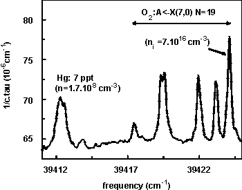
demonstrate the use of CRD for trace gas detection (atomic mercury, known
oscillator strength -> density) and for measuring absolute oscillator strengths
(molecular oxygen, known density -> absorption cross section), a CRD absorption
measurement of ambient air around 253 nm is shown (Figure on the right).
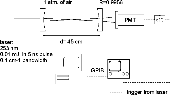
A typical experimental set-up is shown in the figure on the left. The pulsed narrow band laser radiation is usually obtained from a tunable (dye) laser system, that runs at 5-50 Hz, and delivers ns duration pulses with approximately 1-10 mJ of energy. The ring down cavity is formed by two plano-concave mirrors placed at a distance slightly less than twice their radius of curvature apart. The mirrors are coated for an optimum reflectivity in the desired wavelength range, and in the visible and near UV range of the spectrum reflectivities of better than 0.999 up to 0.99999 can be obtained. The mirrors often act at the same time as windows for the closed cell, which contains the substance to be studied. In order to ensure that all transverse modes are detected with equal probability the photo multiplier tube (PMT) that is used to measure the ring down transients is placed directly behind the output mirror. The signal of the PMT is amplified and recorded on a fast and high resolution digitizer. At every wavelength position some pre-defined number of transients, every transient recorded over a sufficiently long time interval, are summed and read out by a PC. The characteristic ring down time is determined by fitting the natural logarithm of the data to a straight line, using a weighted least squares fitting algorithm. Typically, the ring-down time can be determined with a relative accuracy of 10-3. With a typical mirror reflectivity loss of (1-R)=10-4, this implies that line integrated absorption coefficients of better than 10-7, i.e. absorption coefficients below 10-9 cm-1 in a 1 meter cavity, can be measured.
We have presented a pulsed multiplex absorption spectrometer, in which the sensitivity of the Cavity Ring Down absorption detection technique is combined with the multiplex advantage of a Fourier Transform spectrometer. A description of the Fourier Transform - Cavity Ring Down (FT-CRD) spectrometer, substantiated with first experimental results on the atmospheric band of molecular oxygen, is given. It is shown that as in the case of `normal' CRD spectroscopy, the measurement is independent of light intensity fluctuations, provided the spectral intensity distribution of the light source is known and is constant during the measurement (reference).
In a Letter we report on the use of a tunable narrow band continuous laser for sensitive CRD absorption measurements on the weak b1\Sigmag+ (v'=2) <-- X3\Sigmag- (v''=0) band of 18O2. The absorption spectrum is extracted from a measurement of the wavelength dependent Phase Shift (PS) that an intensity modulated cw light beam experiences upon passing through a high Q optical cavity. As the PS-CRD measurement method is independent of random light source intensity fluctuations, it can be performed in a cavity without any stabilization of the cavity length, as is explicitly demonstrated. A comparison of the PS-CRD technique relative to the CRD technique using tunable pulsed light sources is given (reference).
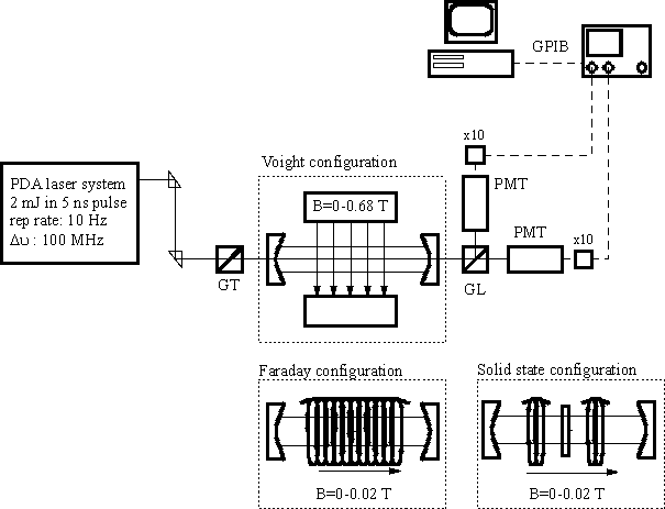
In all the CRD experiments reported to date polarized laser radiation has been used for absorption measurements. By putting a polarization selective optical element in front of the detector, it should in principle be possible to measure the optical rotation of the plane of polarization of the incoming beam upon passage through the ring down cavity. With a polarization analyzer that selects radiation polarized parallel to that of the incoming beam placed in front of the light detector, in addition to the `normal' absorption, rotation of the plane of polarization in the ring down cavity will be detected as an apparent absorption, i.e. as a shortening of the ring down time. With the analyzer rotated (almost) 90 degrees relative to the plane of polarization of the incoming beam, there will still be shortening of the cavity decay time due to absorption but optical rotation will cause a time-dependent increase in the detected signal, i.e. it will appear as an apparent lengthening of the ring down time (`cavity ring up'). When both signals are measured simultaneously, one can discriminate between the effects of absorption and optical rotation. Magnetic rotation spectroscopy \cite{Carroll}, based on the Zeeman effect, is a zero-background absorption technique that is considerably more sensitive than direct single- or double-beam absorption. This technique has been applied to a variety of paramagnetic species, and the resonant magneto-optic spectrum of the $\rm v'=0 \leftarrow v''=0$ transition of the atmospheric band of molecular oxygen has recently been reported \cite{Takubo}. The sensitivity of the technique can in principle be increased by multi-passing through the sample (the polarization rotates further with each traversal). In practice, however, multi-passing is often found to be limited by the deterioration of the degree of polarization with each mirror reflection \cite{Adams,Smith}. We have theoretically outlined and experimentally demonstrated the combination of Polarization Spectroscopy with Cavity Ring Down spectroscopy. The $\rm b^1\Sigma_g^+ (v'=2) \leftarrow X^3\Sigma_g^- (v''=0)$ magnetic dipole transition of molecular oxygen around 628 nm is used to demonstrate the possibility to selectively measure either the polarization dependent absorption or the resonant magneto-optical rotation. The Polarization Dependent CRD (PD-CRD) detection scheme outlined here offers a significant improvement in sensitivity and utility over the individual methods. In contrast to most other schemes for polarization spectroscopy, the PD-CRD scheme is not completely background-free, and is therefore not overly sensitive to the above-mentioned deterioration of the degree of polarization upon multi-passing through the cavity. In the Voigt configuration (external magnetic field perpendicular to the axis of the cavity) the detection limit for molecular oxygen is lowered by more than an order of magnitude in the PD-CRD set-up relative to the `conventional' CRD set-up. In addition, there is a sign-difference for absorptions polarized parallel and perpendicular to the magnetic field, which aids considerably in the assignment. In the Faraday configuration (external magnetic field parallel to the axis of the cavity) the experimental arrangement can be made such that one is exclusively sensitive to optical rotation, and no effect of (polarization dependent) absorption is observed. In the PD-CRD detection scheme, the {\it rate} of polarization rotation is measured, which enables the rotation to be quantitatively determined. The experiments show a noise-equivalent polarization rotation of 10$^{-8}$ rad./cm. Apart from studying electro-optic and magneto-optic phenomena on gas phase species, the PD-CRD technique is shown to be applicable to the study of optical rotation of transparent solid samples as well. (reference).
A simple yet sensitive experimental scheme to record
high-resolution optical absorption and/or optical rotation spectra of molecules
in open air, cells or jets, using ideas from the field of CRD spectroscopy
is demonstrated. Light from a narrow band cw laser is coupled into a high-finesse
stable optical cavity when accidentally coincident with one of the multitude
of modes of this cavity. While rapidly scanning the narrow band laser,
the time-integrated intensity exiting the cavity is monitored. Under the
conditions that the scanning laser is sufficiently long in resonance with
one of the cavity modes that the light intensity inside the cavity approaches
its limiting value, the time-integrated light intensity exiting the cavity
is in a good approximation proportional to the ring down time $\tau$. Direct
absorption spectra and/or optical rotation spectra can therefore be obtained
by plotting the inverse of the time-integrated intensity versus the laser
frequency. With a single mode laser scanning over typically a 1 cm$^{-1}$
spectral region scanning rates of 5-100 Hz have been employed in this study,
yielding high quality spectra in a matter of seconds. The noise equivalent
detection limit readily approaches 10$^{-3}$ of the baseline intensity,
fully comparable to the best CRD spectra reported to date. As the spectral
information is deduced from the time-integrated signal rather than from the
time-dependence of the signal it is possible to perform these measurements with
relatively low power lasers and cheap detection systems. The techniques have
been demonstrated by recording absorption spectra of oxygen, water, and ammonia
in a cell, oxygen and ammonia in a jet, and
magnetic rotation spectra of oxygen in a cell. Furthermore, we have developed a
compact open-path optical ammonia detector, in
which a tunable external cavity diode laser operating at 1.5 mum is used to
probe absorptions of ammonia via the CEA technique.
(reference)
See also our CEA page!
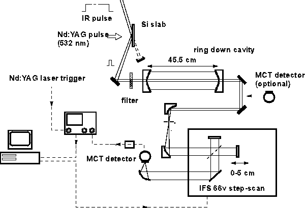
A Cavity Ring Down (CRD) absorption experiment is performed with a Free-Electron Laser (FEL) operating in the 10--11 \mu region. A short infrared pulse of approximately 20 ns, sliced from the much longer FEL pulse, is used to measure CRD spectra of ethylene in two different ways. First, `ordinary' CRD spectra with a resolution determined by the bandwidth of the FEL (\approx 5 cm^{-1}) are recorded. Second, Fourier Transform (FT) CRD spectra with a resolution that is in principle determined by the FT-spectrometer are obtained by analyzing the light exiting the ring down cavity with a FT-spectrometer while the FEL is operated in broad band mode (reference).
The experimental details for the CRD setup do not differ substantially from the setup we have been using before, and a scheme of the experimental setup is shown. The ring down cavity is formed by two identical plano-concave mirrors with a diameter of 25.4 mm and a radius of curvature of -1.0 m placed a distance d = 45.5 cm apart. The reflection coatings are optimized for $\lambda$ = 10.50 $\mu$m, and have a specified reflection coefficient $R \approx $ 0.995. The ring down times thus expected for the empty cavity are on the order of 300 ns. To be able to use the FELIX pulse for CRD experiments it is important to have the trailing flank of the IR pulse decaying faster than the typical photon-lifetime in the cavity. To achieve this, the 'semiconductor switching' technique is applied. This technique is based on the dependence of the reflection and transmission properties of a material on the density profile of free carriers within it. In our case, the second-harmonic radiation of a short-pulse Nd:YAG laser (532 nm, 100 ps, 30 mJ) is used to produce a high density of free carriers in a Si-slab, thereby temporarily changing its IR reflection and transmission characteristics. In the experimental setup, the linearly polarized radiation of the FEL is incident on the semi-conductor Si-slab under Brewster angle ($74 ^{\circ}$ @ 1000 cm$^{-1}$), and passes through the Si-slab in the absence of the Nd:YAG laser pulse. During and shortly after the Nd:YAG laser pulse the Si-slab becomes conducting, and a FEL pulse of approximately 20 ns duration (10 - 20 $\mu$J pulse energy) is efficiently reflected from the Si-slab. This shortened IR pulse is used for the CRD experiments. The IR pulse is coupled into the cavity through one of the mirrors with a weakly focusing mirror (f = 1 m). A long pass filter (7.2 $\mu$m cut-off wavelength) is used to prevent the higher harmonics that might be present in the FELIX-beam from entering the cavity. The time dependence of the light intensity inside the cavity is monitored by detection of the light that leaks out of the cavity through the other mirror. This light is either detected directly or after passage through a FT-spectrometer (Bruker IFS 66v, with step-scan option) using a liquid-nitrogen cooled HgCdTe (MCT) detector (HCT-21-C, Braseby, UK), with a response time of about 40 ns. The signal of the detector is amplified and digitized with a fast (100 Ms/s) and high resolution (10 bit) digital oscilloscope (LeCroy 9430). After averaging of the ring down transients over a pre-defined number of shots (20-50) on the 16 bit on-board memory of the oscilloscope the data are transferred to a PC for extraction of the frequency dependent ring down times.
Rotationally resolved spectra of the b\Sigma (v=0)<-- X\Sigma (v=0)
band of molecular oxygen are recorded by cavity ring down (CRD) spectroscopy
in magnetic fields up to 20 Tesla. Measurements are performed in a 3 cm
long cavity, placed in the homogeneous field region inside a Bitter magnet.
CRD absorption spectra are measured with linearly and circularly polarized
light, leading to different \Delta M selection rules in the molecular transition,
thereby aiding in the assignment of the spectra. Dispersion spectra are
obtained by recording the rate of polarization rotation, caused by magnetic
circular birefringence, using the Polarization Dependent CRD detection
scheme. Matrix elements for the Hamiltonian and for the transition moment
are presented on a Hund's case (a) basis in order to calculate the frequencies
and intensities of the rotational transitions of the oxygen A band in a
magnetic field. All spectral features can be reproduced, even in the highest
magnetic fields. The molar magnetic susceptibility of oxygen is calculated
as function of the magnetic field strength and the temperature, and a discussion
on the alignment of the oxygen molecules in the magnetic field is given.
(click here for more) (click here for pdf file of paper)
A compact diode laser operating around 1.5 micrometer is used to measure absorption spectra of hot water molecules and OH radicals in radiative environments under atmospheric conditions. Spectra of air are measured in an oven at temperatures ranging from 300 K to 1500 K. These spectra contain rovibrational lines from water and OH. The water spectra are compared to simulations from the HITRAN and HITEMP databases. Furthermore, spectra are recorded in the flame of a flat methane/air burner and in an oxyacetylene flame produced by a welding torch. The results show that CEA spectroscopy provides a sensitive method for the rapid monitoring of species in radiative environments.(click here for examples & figures)
We report on the applicability of the Cavity Ring Down (CRD) technique to the study of thin solid films. A measurement of the absorption spectrum around 8.5 micrometer of a 20 -- 30 nm thick C60 film deposited on a 3 mm thick ZnSe substrate, is used to demonstrate that monolayer sensitivity can be obtained. In addition, with the same set-up the transmission losses of the mirrors (about 220 ppm) and the additional losses due to the substrate (less than 340 ppm) can be measured in the same spectral range. (reference)
Cavity Ring Down Spectroscopy / Giel Berden / 2026
Counts since 19/4/2006:
Index molecules [CO] [oxygen, O2] [triphenylamine] [phenol] [acetaldehyde] [dimethylaminobenzonitrile, DMABN]